
Note that the mature forms of the
myeloid series (neutrophils, eosinophils, basophils), all have lobed
(segmented) nuclei.
The degree of lobation increases as the cells mature.

Myeloblast-Promyelocyte-Myelocytes-Metamyelocytes-Band-Segmented
(segs) or polymorphonuclear (PMN)
Myeloblast

|
The earliest recognizable myeloid
cell is the myeloblast (10-20mm diameter) with a large round to oval nucleus. There
is fine diffuse immature chromatin (without clumping) and a prominant
nucleolus.
The cytoplasm is basophilic
without granules. Although one may see a small golgi area adjacent to the
nucleus, granules are not usually visible by light microscopy. One should not
see blast cells in the peripheral blood.
|
|
Promyelocyte 
The promyelocyte (10-20mm diameter) is
slightly larger than a blast. Its nucleus, although similar to a myeloblast
shows slight chromatin condensation and less prominent nucleoli. The
cytoplasm contains striking azurophilic granules or primary granules. These
granules contain myeloperoxidase, acid phosphatase, and esterase enzymes. Under light microscope, usually proomyelocytes are not seen .
At the point in development when
secondary granules can be recognized, the cell becomes a myelocyte.
|
|
Myelocytes

Myelocytes (10-18mm) are slightly
smaller than promyelocytes and have eccentric round-oval nuclei, often
flattened along one side. It has fine chromatin, but shows evidence of
condensation. Nucleoli may be seen in early stages but not in the late
myelocyte. Primary azurophilic granules are still present, but secondary
granules predominate. Secondary granules (neut, eos, or baso) first appear
adjacent to the nucleus. In neutrophils this is the "dawn" of
neutrophilia.
Myelocytes are not normally found
in the peripheral blood.
|
|
Metamyelocytes

Metamyelocytes (10-18mm) are
slightly smaller than myelocytes.
They have kidney shaped indented nuclei and
relatively dense chromatin, especially along the nuclear membrane.
The
cytoplasm is faintly pink with almost no blue background. Numerous secondary granules
(neutro, eos, or baso) clearly outnumber primary granules. Zero to one
percent of the peripheral blood white cells may be metamyelocytes
(juveniles).
|
|
Band

Bands, slightly smaller than
juveniles, are marked by a U-shaped or deeply indented nucleus. Opposite
sides or lobes are of roughly equal size or diameter. There is no nuclear
constriction > than 1/2 the lobe diameter. The chromatin is heavily
clumped and secondary or specific granules either neutrophilic or basophilic
predominate.
Normal band counts vary but are
usually in the range of 0-6%.
|
|
Segmented (segs) or polymorphonuclear
(PMN) leukocytes
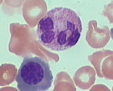
Segmented (segs) or polymorphonuclear
(PMN) leukocytes (average 14 mm dia) are distinguished by definite lobation
with thin thread-like filaments of chromatin joining the 2-5 lobes. The
chromatin of the segmented neutrophil is coarsely clumped and the cytoplasm
is pink due to large numbers of secondary granules.
In practice when examining
peripheral blood, neutrophils are the only leukocytes to be divided into
myelocyte, juvenile, band, and PMN stages. Eosinophils and basophils of all
stages are lumped together in most instances.
Normally approximately 45-75% of
the peripheral blood white cells are segmented neutrophils.
|