Sabtu, 20 Julai 2013

LEFT SHIFT


LEFT SHIFT 


Istilah " left shift " bermakna populasi beberapa sel-sel "beralih" ke arah prekursor belum matang (bermaksud bahawa terdapat lebih prekursor belum matang daripada yang biasa anda lihat). Jika anda lihat siri neutrophil, sebagai contoh. Dalam darah normal, hampir kesemua neutrofil 
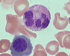
adalah matang (bersegmen). Dalam " left shift ", anda dapat lihat neutrofil yang matang tetapi juga neutrofil yang tidak matang (band, metamyelocytes, myelocytes, dll). Lihat gambar " left shift ", di atas: kebanyakan sel-sel tidak matang.

Rajah : Diagram "Left Shift"


Istilah " left shift " adalah merujuk kepada siri neutrophil. Ianya mula digunakan dahulu apabila pengiraan sel-sel dilakukan secara manual. Kedudukan sel-sel yang paling matang (neutrophil bersegmen) diletakkan untuk butang yang paling kanan, sel-sel kurang matang (myeloblasts) telah diletakkan kepada butang yang paling kiri, dan peringkat sel-sel yang lain terletak diantaranya. Dalam PBF darah yang normal, hampir semua neutrofil terletak di bawah butang paling kanan dalam pengiraan, tetapi kadang-kadang terdapat juga precursor yang awal (contohnya, myelocytes, metamyelocytes, atau promyelocytes). Dalam keadaan ini, sel-sel telah "shifted" ke arah kiri.
Bagi kebanyakan kes, apabila terdapat situasi " left shift ", selalunya pesakit mempunyai jangkitan bakteria. Kadang-kadang " left shift "  juga berlaku apabila terdapat keradangan atau nekrosis.
Berhati-hati, jika anda melihat “nucleated RBC” bersama keadaan " left shift ". Ini dipanggil tindak balas leukoerythroblastotic, ia mungkin menunjukkan masalah yang lebih serius. Kadang-kadang, satu tindak balas leukoerythroblastotic adalah psikologic. Jika hemoglobin adalah sangat rendah (untuk apa jua sebab - kekurangan zat besi yang teruk, kehilangan darah secara besar-besaran), sum-sum tulang akan cuba sedaya upaya untuk menghasilkan sel-sel merah baru dan menghantar mereka keluar ke dalam salur darah secepat mungkin. Keperluan mendesak ini kadang-kadang menyebabkan beberapa prekursor sel darah merah (nucleated RBC) terlepas dari sum-sum tulang. Dan ia juga mula membiarkan prekursor neutrophil (metamyelocytes, myelocytes, promyelocytes) dikeluarkan. Ini adalah tindak balas yang normal kepada kes  anemia yang teruk.
Tindak balas leukoerythroblastotic juga berlaku jika sumsum penuh dengan sesuatu selain tisu hematopoietik - sebagai contoh, carcinoma, atau leukemia - maka sel-sel hematopoietik tidak akan mempunyai ruang yang cukup untuk matang secara sempurna. Sel-sel ini akan keluar meninggalkan sum-sum sebelum mereka matang, dan anda akan melihat kedua-dua sel “nucleated RBC” dan prekursor neutrophil dalam darah. Ini bukanlah sesuatu yang baik..

Figure 1: Haematopoiesis cell development pathways.


Salah satu cara untuk menentukan sama ada tindak balas leukoerythroblastotic  membimbangkan adalah dengan melihat hemoglobin. Jika kandunga hemoglobin adalah sangat rendah (katakan, di bawah 6), maka tindak balas leukoerythroblastotic mungkin adalah secara fisiologi. Walau bagaimanapun, jika hemoglobin adalah normal, atau hanya sedikit menurun, maka tidak ada sebab untuk pesakit untuk mempunyai reaksi leukoerythroblastotic, dan anda perlu lebih memahami apakah penyebabnya

Khamis, 18 Julai 2013

STUDY SLIDE 6 (july)


Question : The blood film of an apparently healthy female aged 20 years who attended a health screening centre where she was found to have a palpable spleen. Hb = 13.7g/dl.‚ red cells 7.21x10^12/l‚ MCV 61.7fl.


Answer : The red cell indices are consistent with a thalassaemia trait. From the Hb and red cell count you will have determined that the MCH is 19pg‚ and this value x100 divided by the MCV gives the MCHC‚ approximately 31g/dl. The red cells are small‚ and in the film many of them show punctate basophilia (Basophilic stippling aka Punctate basophilia refers to an observation found when observing a blood smear in which erythrocytes display small dots at the periphery. These dots are the visualization of ribosomes and can often be found in the peripheral blood smear, even in some normal individuals). The usual explanation of such findings is a thalassaemia. At this age further investigation into the type of thalassaemia may be required and consideration given to future genetic implications.

Isnin, 15 Julai 2013

STUDY SLIDE 5 (JULY)


Question : Comment upon this blood film of a male patient aged 21 years‚ who complains of an acutely swollen and painful knee joint following a recent accident. Hb = 12.3g/dl.‚ red cells 3.76x10^9‚ white cells 140x10^9/l.‚ platelets 737x10^9.


Answer: There are many neutrophils and granulocyte precursors. The latter are mostly myelocytes but there are a few earlier forms‚ which have a high nuclear to cytoplasmic ratio and even at this magnification show prominent nucleoli. This is an example of chronic myeloid leukaemia presenting at an atypically early age. The red cells are not well shown in this picture but you can calculate the MCH

Ahad, 14 Julai 2013

CHEMISTRY : APAKAH "DELTA CHECKS"

DELTA CHECKS

Perbezaan antara keputusan makmal pesakit terkini berbanding dengan keputusan terdahulu yang mana keputusan melebihi paras yang ditetapkan adalah rujuk sebagai delta check. Delta check  disiasat/dikenalpasti secara internal untuk mengenalpasti masalah: 
1) mislabeling, 
2) kesilapan kerani / taip
3) kemungkina kesilapan analisis.
Dalam masa yang sama , panggilan kepada physician atau nurse bertugas perlu dibuat oleh MLT untuk menyiasat kemungkinan kesilapan pre-analytical seperti: 
1) spesimens diambil dari IV lines, 
2) mislabeling, 
3) perubahan sebanar keadaan pesakit.
Pada lapuran makmal pesakit akan dinyatakan tindakan yang telah dilakukan dan spesimen telah diulang kajian sebelum verifikasi. Contoh delta varian ujian chemistry. 

Chemistry (serum)

Albumin 2.0 g/dl
Bilirubin  2.0 mg/dl
*BUN 25 mg/dl
Calcium (Ca) 3.0 mg/dl
Carbon Dioxide 15 mEq/L
Chloride (Cl) 15 mEq/L
*Creatinine 10 mg/dl
Magnesium 2.0 mEq/L
Osmolality  20 mOsm/kg
Potassium (K) 2.5 mEq/L
Sodium (Na) 15 mEq/L
Total Protein 2.0 gm/dl
**Uric Acid 2.0 mg/dl
* Non-Renal
** Non-Heme/Onc

“Autoverification” Keputusan Makmal Klinikal - Apa pendapat anda?


Isu-isu keselamatan pesakit telah menjadi tumpuan masyarakat perubatan . Adakakh Ralat pada keputusa disebabkan oleh kesilapan Manusia. Bagi sebahagian, masyarakat makmal perubatan penyelidukan telah dilakukan untuk mencari jalan cara-cara untuk mengurangkan kesilapan yang dilakukan di dalam makmal.

Salah satu pendekatan ke arah memperbaiki keadaan ini adalah dengan cara mengurangkan penglibatan manusia dalam proses menjalankan ujian dan lebih memilih untuk melakukan kaedah automasi. Bagi hospital atau makmal komersial  yang komprehensif di mana kadar pemprosesan tinggi dan memerlukan pengesahan keputusan ujian yang cepat, autoverification adalah menjadi sebahagian daripada operasi. Dalam makmal yang lebih kecil, autoverification mungkin merupakan langkah pertama yang penting untuk mengurangkan kesilapan manusia dalam laporan keputusan ujian. 

Sabtu, 13 Julai 2013

STUDY SLIDE 4 (JULY)

Question : Comment on this blood film from an elderly male complaining of undue tiredness.


Answer : "The red cells are hypochromic and microcytic. One cell stands out as larger than the others and is stained a ""slate"" blue colour‚ i.e. shows polychromasia‚ the hallmark of a newly released red cell. Iron deficiency anaemia is commonly a symptom of chronic blood loss and this cause always requires consideration before any other. Young red cells inappropriately but universally referred to as reticulocytes complete their maturation in the hours following release into the circulation. There is loss of residual cytoplasmic RNA and consequently loss of basophilic staining. The remains of organelles are removed and the cell membranes are remodelled by the spleen‚ with some decrease in cell size."

Jumaat, 12 Julai 2013

TEKNOLOGI : Bio-Rad Melancarkan The Geenius™ HIV 1/2 Confirmatory System


HERCULES, CA – April 26, 2013 — Bio-Rad Laboratories, Inc. (NYSE: BIO and BIOb),  sebuah syarikat multinational pengeluar dan pengedar produk “life science research” dan “clinical diagnostics”, mengumumkan pelancaran produk “ Geenius™ HIV 1/2 Confirmatory” untuk pasaran antarabangsa.
Geenius™ HIV 1/2 Confirmatory adalah satu sistem yang lengkap menggabungkan ujian HIV-1 and HIV-2 confirmation dan perbezaan dalam serum, plasma atau darah bersama dengan kaedah bacaan automasi dan penterjemahan keputusan yang lengkap - semua dilakukan dalamasa kurang 30 minit.

Faedah
Gabungan dan ujian yang cepat
Pengesahan dan perbezaan  HIV dalamasa kurang 30 minit.
3 jenis sampel (serum, plasma, darah)
Software yang mudah digunakan
Saringan sepenuhnya
 

*tidak perlu bergantung kepada kaedah WESTERN BLOT untuk "HIV confirmatiom".

Khamis, 11 Julai 2013

STUDY SLIDE 3 (july)

QUESTION :

Blood film‚ from an anaemic male patient aged 75 who had a gastrectomy for carcinoma of stomach six months previously. Comment on the findings.


ANSWER :
 
The red cells show marked membrane irregularities‚ principally acanthocytic changes and fragmentation. Haemolysis is sometimes a complication of red cell damage related to metastatic disease.

Isnin, 8 Julai 2013

STUDY SLIDE 2 (july)

Question :
"A film prepared from the blood of a boy who returned to his boarding school following the summer holidays and developed a fever soon afterwards. The provisional diagnosis on the request form was ""?glandular fever"". What do you see?"


Answer : "Two ring forms of Plasmodium falciparum are shown. Routine examination of thick and thin films remains a sound procedure for the diagnosis of malaria despite the development of ""alternative"" methods. The real problem with making the diagnosis is failure to think of it in the first place."

Sabtu, 6 Julai 2013

STUDY SLIDE (july)

QUESTION SLIDE :
Supporting data: A woman of Mediterranean origins aged 30 years‚ the previously healthy mother of three children‚ now complaining of headaches and tiredness. Spleen palpable approximately 4 cm. below the left costal margin. Hb = 9.5 g/dl‚ red cell count 5.0 x10ˆ12/l. Examine the photomicrograph of the patient`s blood film‚ noting your findings and conclusions.


Answer : The red cells are microcytic and hypochromic. A target cell is shown (top left) and there are poikilocytes and small fragmentary forms. The MCH (which you could have obtained from the data provided) is 19 pg. The patient suffered from HbH disease‚ confirmed by electrophoresis and the HbH inclusions test. There was a neutrophil right shift‚ represented in the picture by the cell with six nuclear lobes‚ but a suspicion of folate deficiency was not confirmed by further investigations.

Khamis, 4 Julai 2013

White Cell Basics: Maturation


Note that the mature forms of the myeloid series (neutrophils, eosinophils, basophils), all have lobed (segmented) nuclei. 
The degree of lobation increases as the cells mature.


Myeloblast-Promyelocyte-Myelocytes-Metamyelocytes-Band-Segmented (segs) or polymorphonuclear (PMN)


Myeloblast


The earliest recognizable myeloid cell is the myeloblast (10-20mm diameter) with a large round to oval nucleus. There is fine diffuse immature chromatin (without clumping) and a prominant nucleolus.
The cytoplasm is basophilic without granules. Although one may see a small golgi area adjacent to the nucleus, granules are not usually visible by light microscopy. One should not see blast cells in the peripheral blood.


Promyelocyte


The promyelocyte (10-20mm diameter) is slightly larger than a blast. Its nucleus, although similar to a myeloblast shows slight chromatin condensation and less prominent nucleoli. The cytoplasm contains striking azurophilic granules or primary granules. These granules contain myeloperoxidase, acid phosphatase, and esterase enzymes. Under light microscope, usually proomyelocytes are not seen .
At the point in development when secondary granules can be recognized, the cell becomes a myelocyte.


Myelocytes

 
Myelocytes (10-18mm) are slightly smaller than promyelocytes and have eccentric round-oval nuclei, often flattened along one side. It has fine chromatin, but shows evidence of condensation. Nucleoli may be seen in early stages but not in the late myelocyte. Primary azurophilic granules are still present, but secondary granules predominate. Secondary granules (neut, eos, or baso) first appear adjacent to the nucleus. In neutrophils this is the "dawn" of neutrophilia.
Myelocytes are not normally found in the peripheral blood.

Metamyelocytes


Metamyelocytes (10-18mm) are slightly smaller than myelocytes. 
They have kidney shaped indented nuclei and relatively dense chromatin, especially along the nuclear membrane. 
The cytoplasm is faintly pink with almost no blue background. Numerous secondary granules (neutro, eos, or baso) clearly outnumber primary granules. Zero to one percent of the peripheral blood white cells may be metamyelocytes (juveniles).


Band


Bands, slightly smaller than juveniles, are marked by a U-shaped or deeply indented nucleus. Opposite sides or lobes are of roughly equal size or diameter. There is no nuclear constriction > than 1/2 the lobe diameter. The chromatin is heavily clumped and secondary or specific granules either neutrophilic or basophilic predominate.
Normal band counts vary but are usually in the range of 0-6%.


 Segmented (segs) or polymorphonuclear (PMN) leukocytes


Segmented (segs) or polymorphonuclear (PMN) leukocytes (average 14 mm dia) are distinguished by definite lobation with thin thread-like filaments of chromatin joining the 2-5 lobes. The chromatin of the segmented neutrophil is coarsely clumped and the cytoplasm is pink due to large numbers of secondary granules.
In practice when examining peripheral blood, neutrophils are the only leukocytes to be divided into myelocyte, juvenile, band, and PMN stages. Eosinophils and basophils of all stages are lumped together in most instances.
Normally approximately 45-75% of the peripheral blood white cells are segmented neutrophils.