Selasa, 29 Mac 2022

VDLR vs RPR


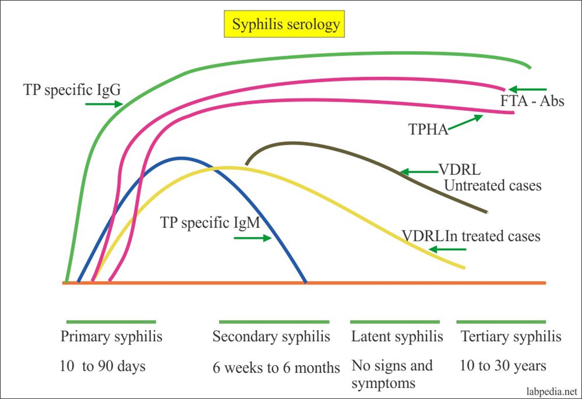
Syphilis is one of the well-known sexually transmitted infections. This disease is caused by the bacteria Treponema pallidum. Syphilis can be detected by different tests. These tests are divided into two parts: the nontreponemal tests and the treponemal tests. 

The nontreponemal tests can detect non-specific treponemal antibodies. There are two common tests under the nontreponemal test. They include VDRL (Venereal Disease Research Laboratory) and RPR (Rapid Plasma Reagin). These two tests are performed in the same way. However, they have a few differences.


VDRL test was developed by the Venereal Disease Research Laboratory during World War I. This test is still done today to detect syphilis.

RPR was developed as a more advanced VDRL. RPR is just the VDRL antigen, but it contains carbon or delicately divided charcoal particles. With these charcoal particles, it allows the visualization of the reaction or flocculation between the specimen and the antigen without the use of a microscope. 


RPR-VDRL (Serologic Tests for Syphilis)

A variety of serologic tests for syphilis are available, including:

1)      VDRL (Venereal Disease Research Laboratory)

2)      RPR (Rapid Plasma Reagin)

3)      FTA-ABS (Fluorescent Treponemal Antibody Absorption)

4)      TP-MHA (Treponema Pallidum Microhemagglutination Assay)

Each differs the others in the precise substance being measured, complexity, and specificity. All are satisfactory for use in managing syphilis. Abnormals may be:

Reactive, Weakly reactive, or Bordeline

Whenever a screening test (RPR, VDRL) is positive, a more specific test (FTA-ABS, TP-MHA) should be used to confirm the test and rule out a "biologic false positive."


A “negative” or "nonreactive" test may indicate:

·         The patient doesn't have syphilis

·         The patient has syphilis, but is so early in the course of the disease that the test has not yet turned positive. In these cases, the test may never turn positive if the patient is effectively treated.

·         The patient had primary syphilis, had a positive test, was effectively treated, 6 months have passed and the test has now reverted to negative.

·         The patient had secondary syphilis, had a positive test, was effectively treated, 12-18 months have passed and the test has now reverted to negative.

·         The patient has syphilis, but his/her immune system is impaired.


A “positive” or "reactive" test may indicate:

·         The patient has syphilis.

·         The patient had syphilis, was effectively treated, but the test has not yet returned to negative:

o   With primarily syphilis, it typically takes about 6 months for the test to turn negative.

o   With secondary syphilis, it typically takes 12-18 months for the test to turn negative.

o   The longer syphilis remains untreated, the longer it will take for the test to return to normal, and the less likely it is to ever return to normal.

·         The patient has a biologic false positive (BFP)


The traditional screening algorithm pathway (see FIGURE 1) relies on the initial analysis of a NTT such as the RPR (rapid plasma reagin) or Venereal Disease Research Laboratory (VDRL) test. If the initial result is reactive, then additional testing is done to confirm the primary result. This additional testing can be done in several ways, including via TP-PA, fluorescent treponemal antibody absorption (FTA-ABS), enzyme immunoassay (EIA), or chemiluminescence immunoassay (CLIA). A reactive result on this confirmatory test is to be considered diagnostic of active T. pallidum infection. Additional specificity for T. pallidum diagnosis is achieved with IgG- and/or IgM-class antibodies detected with the NTT methods described above..

Traditional Testing Algorithm

Non-Treponemal Assay

Treponemal Assay

Interpretation

Non-Reactive

Not Indicated*

No laboratory evidence of syphilis infection

Reactive

Non-Reactive

Syphilis Infection unlikely; biological false positive likely

Reactive

Reactive

Current or past syphilis infection

*If there is high clinical suspicion for early acute disease, then serologic testing should be repeated in 2- 4 weeks, or the patient should be presumptively treated. 


The reverse algorithm (see FIGURE 2) is another alternative used by health care providers. Recent studies have indicated that reverse sequence screening may detect more cases of early or latent syphilis than the traditional forward screening algorithm.6 As recommended by the Centers for Disease Control and Prevention (CDC), discordant syphilis IgG and RPR results are to be resolved by a second treponemal test (TP-PA).7 In the presence of a positive syphilis IgG and non-reactive RPR, a non-reactive treponemal test indicates a false positive syphilis IgG screen because TP-PA has a higher sensitivity than syphilis IgG screening.8 A treponemal confirmation test deemed positive typically results in patient treatment, as the regimen is inexpensive and the consequences of a missed public health opportunity can be large.


This reverse algorithm pathway is gaining traction among obstetric health care providers due to the cross-reactivity of the RPR test with medical conditions such as other infections (eg, HIV), pregnancy, autoimmune disorders, injection-drug use, older age, or immunizations. Screening out this approximately 1% of the population that exists as false positive has become a necessity (see FIGURE 3). Aside from the obvious benefits of lab automation, this method also represents less intensive patient follow-up and stress from treatment. Although not as widely preferred as the forward algorithm, CPL provides guidance to clinicians for interpreting reverse algorithm results (see FIGURE 4). Both of these approaches are summarized in FIGURE 5.


Reverse Testing Algorithm

Treponemal

Assay

Non-Treponemal

Assay

Treponemal

Assay

Interpretation

Non-Reactive

Not Indicated

Not indicated

No laboratory evidence of syphilis infection

Reactive

Non-Reactive

Non-Reactive

Inconclusive for syphilis infection; potentially early infection or false positive. If recent exposure, recommend re-

screening in 2-4 weeks.

Reactive

Non-Reactive

Reactive

Past or potential early syphilis infection

Reactive

Reactive

Not indicated

Current or past syphilis infection


One significant potential limitation of using the reverse algorithm exclusively is the prevalence of the impact of false-positive results that can contribute to a burden of anxiety and treatment.9 However, other more obvious benefits of the reverse screening pathway include the ability to perform the treponemal IgG test on a high throughput analyzer (such as the Bio-Rad BioPlex 2200). Furthermore, the quantitative result obtained from this test aids in the monitoring of disease progression, especially during the tertiary phase. The reverse screening process studied by the University of Iowa Hospitals and Clinics has proven that 99% of results screened negative (1% screened positive).10 While the initial IgG treponemal test may be more expensive per test, overall client satisfaction is improved by providing results more rapidly. 


Jumaat, 15 November 2019

Autoimmune Hepatitis


Diagnosis of autoimmune hepatitis in any patient who has acute hepatitis or acute liver failure (defined by the new onset of coagulopathy include the following assays:
·        Serum antinuclear antibody (ANA)
·        Anti–smooth muscle antibody (ASMA)
·        Liver-kidney microsomal type 1 (LKM-1) antibody
·        Serum protein electrophoresis (SPEP)
·        Quantitative immunoglobulins

Urgent liver biopsy, transjugular if appropriate, may help to confirm the clinical suspicion of autoimmune hepatitis.
Laboratory findings in autoimmune hepatitis include the following:
·        Elevated serum aminotransferase levels (1.5-50 times reference values)
·        Elevated serum immunoglobulin levels, primarily immunoglobulin G (IgG)
·        Seropositive results for ANAs, SMAs, or LKM-1 or anti–liver cytosol 1 (anti-LC1) antibodies
In 50% of patients, abnormal results on liver function tests include decreased albumin levels and prolonged prothrombin time.

Autoantibody Assays
Autoimmune hepatitis is characterized by positive findings on autoantibody tests, as follows:
·        AIH-1 - ASMA and ANA
·        AIH-2 - Anti–LKM-1 antibody
·        AIH-3 - Antibodies to soluble liver antigen (anti-SLA)

+
SMAs are present in 90%-100% of patients with autoimmune hepatitis type 1 (AIH-1). ANAs are present in 10% of patients with AIH-1 and in association with SMAs in 40%-60% of patients with AIH-1. Titers range from 1:100-500,000. SMAs occur in low titers in healthy children and patients with viral hepatitis and other diseases that do not affect the liver.
LKM-1 antibodies are present in 40%-45% of patients with AIH-2 and are associated with anti-LC1 antibodies in 50% of patients. Anti-LC1 antibodies occur alone in 30% of patients with AIH-2; these antibodies recognize formiminotransferase cyclodeaminase, a liver-specific 58kD metabolic enzyme. Anti-asialoglycoprotein receptor antibodies occur more often in patients with AIH-1 and may serve as a marker of inflammatory activity.

Other autoantibodies may be evident. Atypical perinuclear antineutrophil cytoplasmic antibodies (pANCA) are frequently present. Czaja et al have shown that patients with autoimmune hepatitis who have positive test results for actin antibody are younger, more commonly test positive for human leukocyte antigen (HLA)–DR3, and required transplantation more frequently than patients with ANAs who test negative for actin antibody

Serum Proteins and Immunoglobulins
An IgG-predominant polyclonal hypergammaglobulinemia is a common finding in patients with untreated autoimmune hepatitis. Gamma globulin values typically range from 3-4 g/dL and frequently are as high as 5-6 g/dL. Cases of hyperviscosity syndrome secondary to high IgG levels are reported. Autoimmune hepatitis is an unlikely diagnosis in patients who have acute hepatitis without hypergammaglobulinemia.
The gamma globulin or the IgG level may be followed on a regular basis as a marker of disease responsiveness to therapy.
Patients with AIH-2 commonly have partial immunoglobulin A (IgA) deficiency.

Aminotransferases
Serum aminotransferases (aspartate aminotransferase [AST] and alanine aminotransferase [ALT]) are elevated in 100% of patients at initial presentation, with average values of 200-300 U/L. Aminotransferase values correlate poorly with the degree of hepatic necrosis; however, values in the thousands may indicate acute hepatitis or a severe flare of preexisting disease.
Continued elevation of the aminotransferases in the face of continuing therapy is a reliable marker for ongoing inflammatory activity in the liver. Normalization of the aminotransferase levels during therapy is an encouraging sign, but active liver inflammation is present in more than 50% of patients with normalized liver chemistries. Indeed, biochemical remission may precede true histologic remission by 3-6 months.
Typically, patients are treated for at least 1 year after documentation of normal liver chemistries. Liver biopsy is recommended by some experts to confirm that the patient is in histologic remission. Drug withdrawal may be attempted at this time (see Treatment).

Worsening of aminotransferase levels in a patient undergoing treatment or in a patient who is in remission may signal a resurgence of disease activity.

Other liver chemistries
Serum bilirubin and alkaline phosphatase values are mildly to moderately increased in 80%-90% of patients. A sharp increase in the alkaline phosphatase values during the course of autoimmune disease might reflect the development of PSC or the onset of hepatocellular carcinoma as a complication of cirrhosis.
Hypoalbuminemia and prolongation of the prothrombin time are markers of severe hepatic synthetic dysfunction, which may be observed in active disease or decompensated cirrhosis.

Complete Blood Count and Other Blood Studies
Other hematologic abnormalities may include the following:
·        Mild leukopenia
·        Normochromic anemia
·        Coombs-positive hemolytic anemia
·        Thrombocytopenia
·        Elevated erythrocyte sedimentation rate
Eosinophilia is uncommon, but counts ranging from 9% to 48% are described. Autoimmune hepatitis has even been described as the sole presenting feature of idiopathic hypereosinophilic syndrome.

Hepatic Imaging Studies
Imaging studies, in general, are not helpful in reaching a definitive diagnosis of autoimmune hepatitis; however, the presence of heterogeneous hepatic echotexture on abdominal ultrasound or abnormal contrast enhancement on abdominal CT imaging may suggest the presence of active inflammation or necrosis.

The appearance of an irregular nodular liver may confirm the presence of cirrhosis. Furthermore, these imaging studies may be used to rule out the presence of hepatocellular carcinoma, a potential complication of autoimmune hepatitis–induced cirrhosis.

When alkaline phosphatase levels are 7-8 times reference values or gamma glutamyl transferase levels are 2-3 times reference values, a patient with autoimmune hepatitis and ulcerative colitis may require endoscopic retrograde cholangiopancreatography (ERCP) to rule out coexisting primary sclerosing cholangitis (PSC).

Liver Biopsy
Liver biopsy is the most important diagnostic procedure in patients with autoimmune hepatitis. This procedure can be performed percutaneously, with or without ultrasound guidance, or by the transjugular route. The latter is preferred if the patient has coagulopathy or severe thrombocytopenia. A transjugular liver biopsy also may be preferable if ascites is present or if the liver is small, shrunken, and difficult to reach percutaneously.

Liver biopsy routinely is performed in the outpatient setting to investigate abnormal liver chemistries. Liver biopsy should be performed as early as possible in patients with acute hepatitis who are thought to have autoimmune hepatitis. Confirmation of the diagnosis enables initiation of treatment at an early stage in the disease process.

The role of biopsy in patients presenting with well-established cirrhosis secondary to autoimmune hepatitis is less clear. As an example, the initiation of treatment in a patient with cirrhosis, normal aminotransferase levels, and a minimally elevated gamma globulin level is not expected to influence the disease outcome.


Histologic Findings
Histopathologic findings on liver biopsy specimens are crucial to determining the diagnosis of autoimmune hepatitis and the disease's severity. Liver biopsy findings can help to differentiate autoimmune hepatitis from chronic hepatitis C virus (HCV) infection, alcohol-induced hepatitis, drug-induced liver disease, primary biliary cirrhosis, and PSC. [43]

Autoimmune hepatitis is characterized by a portal mononuclear cell infiltrate that invades the limiting plate surrounding the portal triad and permeates the surrounding lobule (ie, periportal infiltrate) and beyond. A plasma cell infiltrate sometimes occurs, which, in the past, led to the use of the term plasma cell hepatitis.

Biopsies may show evidence for interface hepatitis (ie, piecemeal necrosis), bridging necrosis, and fibrosis. Interface hepatitis essentially spares the biliary tree but may involve most of the lobule. Lobular collapse, best identified by reticulin staining, is a common finding.

Interface hepatitis does not predict a progressive disease course. By contrast, a strong likelihood exists that cirrhosis will develop when bridging necrosis is present. The presence or absence of cirrhosis on liver biopsy is an important determinant of the patient's prognosis.

Fibrosis is present in most patients with autoimmune hepatitis. Without effective therapy, fibrosis starts to connect the portal and central areas, which ultimately leads to cirrhosis.

In 1999, the International Autoimmune Hepatitis Group established a scoring system that is particularly helpful in establishing the diagnosis of autoimmune hepatitis in problematic cases. [44, 45]

Histopathologic findings in patients with autoimmune hepatitis are characteristic but nonspecific; autoimmune hepatitis has findings in common with chronic viral hepatitis, drug-associated chronic hepatitis, and several other chronic liver disorders. Multinucleated giant hepatocytes are found in 10%-20% of biopsy specimens; their occurrence after the neonatal period may suggest a diagnosis of autoimmune hepatitis.

Tucker et al indicate that the presence of characteristic hyaline droplets in the cytoplasm of Kupffer cells on routine hematoxylin and eosin (H&E) from biopsy specimens in pediatric patients with autoimmune hepatitis may provide a useful diagnostic clue to distinguish this disease from other forms of chronic hepatitis.


Credit to WebMD LLC. 

Sabtu, 17 Disember 2016

SMEAR SEL DARAH NORMAL

Sel darah merah atau eritrosit, Eritrosit berasal dari bahasa Yunani yakni Erythros (merah) dan kytos (selubungi sel darah). Sel darah merah ( eritrosit ) berbentuk bulat pipih dan cekung pada bagian tengahnya ( biconcave ). 
Pic1 - RBC bentuk 3D

Eritrosit berwarna merah karena mengandung hemoglobin. Sel darah merah ini dibentuk dalam sumsum tulang, kemudianya beredar ke seluruh bagian tubuh yang melalui pembuluh darah. 

Jangkahayat sel darah merah lebihkurang 120 hari. Sel darah merah yang telah tua akan dimusnahkan pada organ hati dan limpa. Sebelum anda benar-benar boleh mempelajari perubahan patologi dalam sel-sel merah, anda perlu tahu bentuk morfologi normal sel darah merah. Berikut adalah imej smear sel darah yang normal. Terdapat beberapa perkara yang perlu perhatikan di sini.
Pic1.1 Smear Normal RBC

Pertama, sel-sel tersebar samarata di lapangan. Ada yang bersentuhan dan ada yang kelihatan sedikit bertindih, sel-sel tidak semua bertindihan di atas satu sama lain. 
Terdapat sedikit ruangan diantara sel-sel tersebut, tiada ruangan/kawasan yang luas diantara sel-sel merah. 
Ini menunjukkan fokus berada dalam kawasan smear darah yang sesuai untuk dibuat penilaian. Fokus kawasan kajian yang betul - "zon morfologi" - adalah terletak pada bahagian tengah smear (diantara bahagian ekor dan kepala). 
Dalam zon ini, sel-sel hampir tidak menyentuh diantara satu sama lain, seperti yang dilihat dalam foto ini.
Pic2 : Smear Normal RB

Kedua, (rujuk Pic2) sel-sel kelihatan mempunyai jumlah hemoglobin yang normal . 
Lihat kawasan putih di tengah-tengah setiap sel merah. Itulah "zon pucat pusat." Ia meliputi kira-kira 1/3 diameter keseluruhan sel merah  (sel-sel seperti ini dipanggil normochromic). 
Jika ia lebih besar, ini bermakna bahawa sel tidak mempunyai hemoglobin yang mencukupi (sel seperti ini dipanggil hypochromic) dan dikenali sebagai anemia.

Seterusnya, lihat bentuk sel-sel ini: semuanya berbentuk bulat. Itulah apa morfologi sel yang normal. Secara keseluruhan, tidak ada sejumlah besar sel-sel merah yang berbentuk (seperti bentuk runcing, bentuk titisan air mata, atau yang berbentuk sabit).

Kepelbagaian bentuk seperti ini dipanggil poikilocytosis (rujuk Pic3). 
Darah yang normal mempunyai poikilocytosis yang minimum (kebanyakan sel-sel darah berbentuk sempurna dan bulat). Sesetengah kes anemia, seperti kes thalassemia yang teruk, mempunyai poikilocytosis yang ketara (kebanyakan sel mempunyai kepelbagaian bentuk).

Pic3 : Poikilocytosis

Pic3.1  : Lebih banyak variasi dan saiz sel darah (anisopoikilocytosis),  Nilai RDW lebih tinggi.

Pic3.2 : Antara variasi sel darah

Pic3.2.1 : Basophilic stippling


Pic3.2.2 : Kes Thalassaemia dengan Basophillic stippling


Pic3.2.3 RBC dengan basophilic stippling/basophilia (sel darah merah yang mengandungi satu titik kecil pada bahagian tepi dinding sel). Ia adalah granul RNA atau gumpalan ribosom. Kadang-kadang terlihat pada smear yang normal. Mudah dilihat pada pesakit yang mengalami “lead poisoning”.


Pic3.2.4


Pic3.2.5 Nucleated RBC - Nucleated red cells - Sel darah dengan nukleus padat dan berwarna gelap di tengah-tengah sel. Dapat dilihat pada bayi (3-4 hari) . Kronik haemolytic anemia. Megaloblastic anemia. Jangkitan kongenital (syphilis, CMV, rubella). Postspleenectomy. Tindakbalas Leukoerythroblastic. Jangkitan Fungal dan mycobacterial. Dyselectropoeitic anemia


Sukar untuk mengukur secara visual saiz sel darah merah. Di bawah mikroskop, agak sukar untuk menentukan sama ada saiz sel-sel darah merah adalah bersaiz normal (normocytic), bersaiz kecil (microcytic) (Pic4), atau bersaiz besar (macrocytic) (Pic4.1). 
Anda perlu melihat nilai MCV (Mean corpuscular volume), nilai ini menentukan sama ada sel-sel darah mempunyai saiz yang normal atau tidak. 

MCV normal adalah diantara 80 dan 100 femtoliters; sel darah merah dengan MCV kurang daripada 80 dianggap microcytic (sel darah bersaiz kecil), dan sel-sel merah dengan MCV lebih 100 dianggap macrocytic (sel darah bersaiz lebih besar). Ini adalah kriteria penting untuk menilai kes anemia 

Pic4 : Normal dan Microcytic sel


Pic5 : Normal dan Macrocytic sel.

Oleh itu, dengan pengamatan menggunakan mikroskop adalah mungkin untuk mengetahui, berapa banyak sel-sel darah yang berbeza dari segi saiz dan bentuk. 
Anda boleh lihat, dengan mata kasar anda, sama ada sel-sel darah mempunyai banyak saiz yang sama, atau sama ada terdapat pelbagai saiz dari yang kecil kepada saiz yang besar. 

Sel darah yang normal biasanya bersifat anisocytosis yang minimum (purata sel-sel darah bersaiz sama), beberapa kes anemia, seperti anemia kekurangan zat besi yang teruk, keadaan anisocytosis adalah lebih ketara (terdapat banyak sel darah yang kecil dan yang besar).

Parameter CBC yang menunjukkan variasi dalam saiz sel adalah nilai RDW. Jika nilai RDW adalah rendah, ini bermakna keadaan anisocytosis adalah minimum, dan kebanyakan sel mempunyai saiz yang hampir sama. Jika RDW adalah sangat tinggi, bermakna terdapat anisocytosis yang ketara, dimana sel-sel berbeza dari segi saiz. Ini adalah satu lagi nilai yang boleh membantu anda apabila anda cuba untuk mendiagnosis kes anemia.


Pic5 Kes IDA yang teruk. Pelbagai saiz dan morfologi sel dapat dilihat.


Akhir sekali, bagi satu smear darah yang normal, sepatutnya anda tidak akan menjumpai “nucleated red blood cells”, organisma berjangkit (seperti organisma malaria) dalam sel-sel merah, “Pappenheimer bodies”,  “Howell-Jolly”  atau apa saja morfologi yang pelik.
Pic6  : Howell-Jolly bodies – Adalah partikel intracellular berbentuk bulat (kromatin DNA yang padat). Biasanya terdapat hanya satu pada sel, tetapi kadang-kadang boleh lebih dari satu. Tardapat pada kes Megaloblastic anaemia dan Mylodysplastic

Pic6.1 : Howell-Jolly bodies

Pic6.2 : Pappenheimer body - Granul abnormal iron yang terdapat dalam sel darah. Granul blue-purple yang padat.  Terdapat pada individu yang mengalami sideroblastic anemia, hemolytic anemia, dan penyakit sickle cell.

Pic6.3 : Pappenheimer body

Pic6.4 : Pappenheimer body


Jumaat, 6 November 2015

Ujian Saringan Dadah Dalam Air Kencing

Ujian Saringan Dadah Dalam Air Kencing


 PELAKSANAAN KOD AMALAN PENCEGAHAN DAN PEMBASMIAN PENYALAHGUNAAN DADAH, ALKOHOL DAN BAHAN DI TEMPAT KERJA dan Ujian Saringan Dadah Dalam Air Kencing.


Kod Amalan ini bertujuan membantu majikan dan pekerja bagi memenuhi tanggungjawab di bawah Akta Keselamatan dan Kesihatan Pekerjaan (AKKP) 1994 untuk menangani masalah dadah, alkohol dan bahan di tempat kerja. Di bawah Seksyen 15 AKKP, majikan bertanggungjawab untuk memastikan keselamatan, kesihatan dan kebajikan semua pekerjanya. Seksyen 24(1) AKKP pula menjelaskan bahawa pekerja bertanggungjawab untuk memberi perhatian yang munasabah bagi keselamatan dan kesihatan dirinya dan orang lain yang mungkin terjejas oleh tindakannya di tempat kerja. Untuk itu, Bahagian Kesihatan Pekerjaan telah dipertanggungjawab bagi melaksanakan pemantauan pematuhan Koda Amalan Pencegahan dan Pembasmian Penyalahgunaan Dadah, Alkohol dan Bahan Di Tempat Kerja di peringkat negeri dengan cara melakukan Ujian Saringan Dadah Dalam Air Kencing. Majikan atau tempat kerja dipantau dipandu pada beberapa aspek, seperti mengadakan dasar, pembentukan Jawatankuasa Pencegahan dan pembasmian penyalahgunaan dadah, alkohol dan bahan di tempat kerja dan latihan pekerja dan pegawai-pegawai yang bertanggung jawab.

Antara jenis-jenis dadah dalam Ujian Saringan Dadah Dalam Air Kencing adalah :